
Single molecule and cell biophysics for Biomedicine - Hatzakis Lab
The main objective of my group is to augment our understanding on the molecular mechanisms that underlie and control vital cellular functions. We approach this challenge by deciphering the dynamic interplay between the function and spatiotemporal localization of biomolecules (virus, dug nanocarrier, oligonucleotides or protein assemblies) and how this correlate to cellular and organismal response.
Novel new imaging technologies
We have therefore pioneered the development of novel new imaging technologies - with an emphasis on single particle and live cell microscopy - that promises to shed light on the interplays between the behaviour (dynamics, function and localisation) of biomolecules and high throughput single particle screening methodologies to decipher oligonucleotide interactions with membranes. By tracking the spatiotemporal displacement and localization of one protein - or an assembly - in real time we revealed internalization pathways and cell fate of nanoparticles and viruses.
Our functional and FRET studies at the fundamental limit of individual catalytic cycle, on the other hand have helped deciphering protein structure and function dependence on temporal cell localization and the design of novel ligand biasing aberrant biological function.
Recognizing that 4D imaging generates terabytes of data sets, that are prohibitively hard to be quantitatively evaluated by current semi-manual analysis, we have developed toolboxes and softwares based on machine learning to rapidly, reliably and free of human cognitive biases, analyze the wealth of novel microscopy data we, and others, produce. Our all-inclusive softwares for windows and macs, offer transition from raw data to quantitative analysis and data classification with <5 clicks, accelerating the automatic analysis by ~6 orders of magnitude.
These combined methodologies bridge 4D imaging with sophisticate image analysis required for delving into the era of 4D cell and tissue imaging.
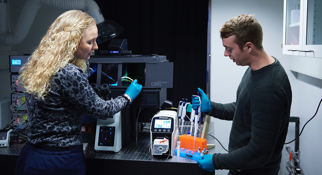
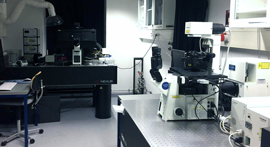
The Hatzakis Group labs are equipped with everything needed to perform state of the art experiments on a variety of protein, enzyme and related experiments. Most experiments in the group are build around fluorescent microscopy, performed on one (or both) of two microscopes:
- An IX81 Olympus confocal microscope
- An Olympus Total Internal Reflection Fluorescence microscope (TIRFm) set up for super resolution microscopy
- State of the art Olympus SpinSR10 Confocal Super Resolution Microscope
- GMO facilities: for cell culture and protein expression and purification
Accelerating biological discoveries by machine learning and quantitative single particle microscopy
Advanced microscopic techniques produce vast amounts of unstructured data the analysis of which by conventional methodologies is tedious, time consuming and may be biased by unconscious biases. We have been developing agnostic quantitative and automated analysis methodologies based on machine learning to treat classify and annotate biological behaviors. The toolboxes offer rapid analysis often accelerated by 3- 6 orders of magnitude, help eliminating potential human biases and provide statistical insights on biological parameters that underlie and control protein function and cellular responses.
Relevant publications
- Jacob Kæstel-Hansen et al. Deep learning-assisted analysis of single-particle tracking for automated correlation between diffusion and function. Nat Methods 22, 1091–1100 (2025).
- Steen W. B. Bender et al. SEMORE: SEgmentation and MORphological fingErprinting by machine learning automates super-resolution data analysis. Nat Commun 15, 1763 (2024).
- Malle, M. G., et al. Single-particle combinatorial multiplexed liposome fusion mediated by DNA. Nature Chemistry (2022) 14, 558-565
- Pinholt, H. D. et al. Single Particle Diffusional Fingerprinting A machine learning framework for quantitative analysis of heterogeneous diffusion.PNAS (2021), 31, 118.
- Thomsen, J., et al. DeepFRET, a software for rapid and automated single- molecule FRET data classification using deep learning. eLife (2020), 9.
High throughput single-nanocontainer readouts and deep learning: Pushing the biomolecular recognition detection to new frontiers
Screening of biomolecular recognition often suffers from challenges such as long running time, high person power as well as excessive materials cost. To surpass these challenges have developed miniaturized assays for ultra-sensitive and high-throughput screening of biomolecular interactions and to explore:
- DNA-DNA recognition and sub-attolitter cargo delivery,
- Transporter function and
- How membrane properties affect protein function.
Rapid and reliably analyis and classification of the multidimensional multi terabyte data is achieved by our deep learning analytic tools.
Relevant publications
- Malle, M. G., et al. Single-particle combinatorial multiplexed liposome fusion mediated by DNA. Nature Chemistry (2022) 14, 558-565
- Schmidt, S. G., et al. The dopamine transporter antiports potassium to increase the uptake of dopamine. Nature Communications(2023) 13, 2446
- Thomsen, S. P., et al. A large size-selective DNA nanopore with sensing applications. Nature Communications (2019) 175, 5655
Metabolic pathways redefined: Biased P450 metabolism by smart ligands targeting protein dynamics and targeting metabolic diseases
POR is a central molecular hub activating a plethora of metabolic pathways by donating electrons to more than 50 different cytochrome P450 enzymes (CYPs). Point mutations in POR cause severe metabolic disorder due to altered POR-CYP interactions. In collaboration with Paediatric Endocrinology at the University Hospital Bern, Switzerland, and Department of Plant Biology, University of Copenhagen, we study these interactions all the way from clinical phenotype down to the fundamental limit of individual proteins. Combining single molecule FRET and single turnover studies with cell studies and docking simulations we advance our understanding on the intricate role of conformational dynamics to activity and specificity and eventually how pathogenic mutations and small molecule ligand interactions control metabolic disorder and biosynthetic pathways.
Relevant publications
- Jensen, S.B. et al. Biased cytochrome P450-mediated metabolism via small-molecule ligands binding P450 oxidoreductase. Nature Communications(2021), 12, 2260.
- Laursen, T. et al. Characterization of a Dynamic Metabolon Producing the Defense Compound Dhurrin in Sorghum. Science (2017), 354, 890-893.
- Bavishi, K. et al. Direct Observation of Multiple Conformational States in Cytochrome P450 Oxidoreductase and their Modulation by Membrane Environment. Scientific Reports (2018), 8, 1-9.
- Laursen, T. et al. Single Molecule Activity Measurements of Cytochrome P450 Oxidoreductase Reveal the Existence of Two Discrete Functional States. ACS Chem. Biol. (2014), 9, 630-634.
Deciphering cellular choreography: Insights into single particle of proteins, viruses, and pharmaceutics nanocarriers
We have pioneered the development of powerful methodologies to track the spatiotemporal localization in live cells of individual biomolecules,(proteins organelles viruses and nanocarriers) and quantify their interaction with membranes cell entry pathways and utilized this information to tailor their targeted delivery directly. To analyse the complex, multidimensional, multiterabyte data we acquire, we have employed novel methodologies based on machine learning that offer rapid precise and automated transition from raw microcopy images to quantitative biomedicine insights accelerating discoveries often by 104 times.
Relevant publications
- Jacob Kæstel-Hansen et al. Deep learning-assisted analysis of single-particle tracking for automated correlation between diffusion and function. Nat Methods 22, 1091–1100 (2025).
- Anwesha Sanyal et al. Neuronal constitutive endolysosomal perforations enable α-synuclein aggregation by internalized PFFs. J Cell Biol (2025) 224 (2): e202401136.
- Pinholt, H. D. et al. Single Particle Diffusional Fingerprinting A machine learning framework for quantitative analysis of heterogeneous diffusion.PNAS (2021), 31, 118.
- Moses E. M.,et al., ACS Applied Materials & Interfaces (2021) 13 (28), 33704- 33712
- Wan, F., et al. Ultrasmall TPGS–PLGA Hybrid Nanoparticles for Site-Specific Delivery of Antibiotics into Pseudomonas aeruginosa Biofilms in Lungs. ACS Appl. Mater. Interfaces (2019) 12, 1, 380–389
Bridging structure and function of CRISPR-Cas12a with smFRET and Cryo-EM
Adaptive immunity in bacteria is accomplished by the CRISPR system, and CRISPR- associated proteins (Cas). Proteins coupled with RNA are guided by this system to recognize and cleave foreign genetic material. As such, it’s also a powerful method for genome editing, and is receiving lot of bio technical and medical attention currently. By using single molecule FRET we can study this system in great detail, and obtain a wealth of structural and kinetic information, when combining with other techniques. Read how we did this in Stella et al. (2018), published in Cell.
Relevant publications
- Stella, S., et. al. Conformational Activation Promotes CRISPR-Cas12a Catalysis and Resetting of the Endonuclease Activity. Cell (2018), 175, 1856–1871.e21
- Thomsen, J., et al. DeepFRET, a software for rapid and automated single-molecule FRET data classification using deep learning. eLife (2020), 9
- NanoPANS project funded by the Lundbeck Foundation (2025)
- NovoNordisk foundation Challenge Center for Optimized oligo Escape and control of disease (2024)
- The 4D Cellular Dynamics (2023)
-
2014: Villum Foundation, Young Investigator Fellowship

-
2016: Novozymes A/S & The Henning Holck-Larsen Foundation, Guest Post-doctoral Fellowship
-
2016: Novo Scholarship Programme

-
2017: Carlsberg Foundation, Most Distinguished Associate Professor Fellowship
![]()
-
2017: Marie Curie, Post Doc Fellowship

-
2017: Lundbeck fonden, Post Doc Fellowship

-
2017: Velux foundation Center: Advanced Biomolecular Engineering

-
2018: Innovation Foundation Denmark, Industrial Post-doc

DeepFRET
Rapid and automated single molecule FRET data classification using deep learning.
Extraction of liposome intensity
Python based script for extraction and analysis of .tif formatted image files. The script can extract the intensity from individual liposomes based in a changeable ROI size given initially and subtract local background.
Cell analyzer HEK293/PYY/eYFP
Python based script for extraction and analysis of .tif formatted image files. The script will identify cells on a image based on set thresholds and parameters. From this a mask will be created to extract the intensity from up to three channels, here corresponding to signal from membrane stain (blue), yellow fluorescent protein (YFP, green) and Cy5/Atto655 (red).
Single Particle Tracking of Lipases
Python Script for analysing .tif movies of lipases diffusing on a surface. Code used to make data for Bohr, S. S.-R. et al. Scientific Reports, 2019.
Diffusional fingerprinting
An all inclusive tool for SPT data analysis, processing, and classification. The method uses machine learning for dissecting the features that underlie diffusional behavior and establishing molecular identity, regardless of the underlying diffusion type. See our recent paper in PNAS 2021.
https://github.com/hatzakislab/Diffusional-Fingerprinting
Staff
| Name | Title | Phone | |
|---|---|---|---|
| Aimilia Nousi | Special Consultant | +4535333053 | |
| Artu Breuer | Assistant Professor | +4535326895 | |
| Athanasios Oikonomou | PhD Fellow | ||
| Frank Høgh Schulz | Postdoc | +4535323121 | |
| Freja Schmidt-Rasmussen Bohr | PhD Fellow | ||
| Georgios Bolis | PhD Fellow | +4535328401 | |
| Georgios Kyriakakis | PhD Fellow | ||
| Janni Støvring Herzberg | Postdoc | ||
| Konstantinos Tsolakidis | Academic Research Staff | +4535328863 | |
| Marcus Winther Dreisler | PhD Fellow | ||
| Min Zhang | Assistant Professor | ||
| Nikos Hatzakis | Professor | +4535334502 | |
| Richard Michael | PhD Fellow | ||
| Sara Vogt Bleshøy | Postdoc | +4535326121 | |
| Stavroula Margaritaki | PhD Fellow | +4535321061 | |
| Steen Wielandt Barfod Bender | PhD Fellow | ||
| Tania Sabina Darphorn | Special Consultant | +4593565147 | |
| Victoria Jade Kladny | PhD Fellow | +4535324958 |
Master Student
| Name | Title | Phone | |
|---|---|---|---|
| Emilie Elisabeth Milan Nielsen | Master Student | ||
| Kterina Vougiatzi | Master Student | ||
| Freya Rerihold | Master Student | ||
| Sascha Valetin Brown | Master Student | ||
| Athanasia Vapori | Master Student |
Bachelor Student
| Name | Title | Phone | |
|---|---|---|---|
| Molly Jean Maud Turner | Bachelor Student | ||
| Julie Bernt Frederiksen | Bachelor Student | ||
| Josefine Bjørcklind | Bachelor Student |
Contact

Nikos Hatzakis
Professor, group leader
Nano Science Center
Department of Chemistry
Office: T554
Phone: +45 50 20 29 51
E-mail: hatzakis@chem.ku.dk
Orcid
https://orcid.org/0000-0003-4202-0328
Center for Optimized Oligo Escape and Control of Disease (COE)